
Image-guided stereotactic radiotherapy (SRT) combines precise radiation delivery with advanced imaging, enabling accurate tumor targeting and real-time adjustments for optimal treatment outcomes in various cancers․
1․1 Definition and Overview
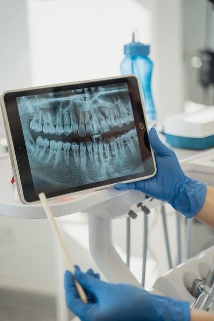
Image-guided stereotactic radiotherapy (IG SRT) is a highly precise radiation therapy technique that uses advanced imaging technologies to accurately target tumors while minimizing damage to surrounding healthy tissues․ By integrating real-time imaging with sophisticated radiation delivery systems, IG SRT ensures accurate positioning and dose delivery, even for complex or moving tumors․ This approach is particularly effective for treating intracranial and extracranial tumors, offering non-invasive alternatives to traditional surgical methods․ The core principle of IG SRT lies in its ability to adapt treatment plans dynamically, leveraging technologies like cone-beam CT, MRI, or robotic systems to achieve superior clinical outcomes․ Its applications span various cancers, including brain, lung, liver, and prostate tumors, making it a cornerstone of modern radiation oncology․
1․2 Historical Development
The evolution of image-guided stereotactic radiotherapy (IG SRT) began with the introduction of stereotactic techniques in the 1950s, initially focused on intracranial tumors․ Early systems relied on rigid frames and basic imaging, limiting precision․ The 1980s saw advancements with computed tomography (CT) integration, enabling better tumor localization․ The 1990s marked the emergence of image-guided radiation therapy (IGRT), combining stereotaxy with real-time imaging modalities like cone-beam CT (CBCT) and MRI․ This integration revolutionized SRT, allowing for precise targeting without invasive frames․ The 2000s introduced robotic systems, such as CyberKnife, further enhancing accuracy and adaptability․ Recent advancements incorporate artificial intelligence and adaptive radiotherapy, improving efficiency and patient outcomes․ This historical progression underscores the transformation of SRT into a highly sophisticated, non-invasive cancer treatment modality․
1․3 Importance in Modern Radiation Oncology
Image-guided stereotactic radiotherapy (IG SRT) plays a pivotal role in modern radiation oncology due to its unmatched precision and effectiveness․ By integrating advanced imaging technologies, IG SRT ensures accurate tumor targeting, minimizing exposure to surrounding healthy tissues․ This is particularly crucial for treating tumors in sensitive areas, such as the brain, prostate, and lungs․ The ability to deliver high doses in fewer fractions enhances treatment efficiency and patient comfort․ Additionally, IG SRT’s adaptability to tumor motion and anatomical changes allows for real-time adjustments, improving outcomes and reducing side effects․ Its non-invasive nature makes it an attractive alternative to surgery for many patients․ Consequently, IG SRT has become a cornerstone in contemporary radiation oncology, offering personalized and effective cancer care․

Principles of Image-Guided Radiation Therapy (IGRT)
IGRT combines advanced imaging modalities with precise radiation delivery, enabling real-time tumor localization and adjustments to ensure accurate and effective treatment while minimizing damage to surrounding tissues․
2․1 Imaging Modalities in IGRT
In IGRT, various imaging modalities such as Cone-Beam CT (CBCT), MRI, PET, and 4D-CT are employed to enhance precision․ CBCT provides real-time 3D imaging, integrated with linear accelerators, allowing accurate patient positioning and tumor targeting․ MRI offers superior soft-tissue contrast, beneficial for tumors in organs like the liver or prostate․ PET scans highlight metabolically active tumor regions, aiding in precise dose delivery․ 4D-CT captures tumor motion over time, essential for treating moving targets such as lung tumors․ These modalities enable dynamic adjustments, ensuring radiation is delivered accurately while sparing healthy tissue․ The choice of imaging modality depends on tumor location, size, and patient-specific factors, ensuring optimal treatment outcomes․
2․2 Target Localization and Tracking
Image-guided SRT employs advanced techniques for precise target localization and tracking, ensuring accurate radiation delivery․ Fiducial markers or implanted sensors are often used to identify tumor locations, particularly in moving organs like the prostate or liver․ Real-time tracking systems, such as optical surface guidance or electromagnetic transponders, monitor tumor motion during treatment․ For lung tumors, 4D-CT captures movement linked to breathing, enabling synchronized radiation beams․ These methods enhance targeting accuracy, reducing margins and sparing healthy tissue․ Dynamic adjustments allow for precise dose delivery, even for tumors in challenging locations․ This integration of imaging and tracking ensures that radiation is focused on the target, improving outcomes and minimizing side effects in stereotactic radiotherapy․
2․3 Real-Time Adjustments for Precision
Real-time adjustments in image-guided SRT enhance treatment precision by correcting for anatomical changes or tumor motion during delivery․ Advanced imaging systems, like cone-beam CT or MRI, provide immediate feedback, allowing clinicians to adapt treatment plans dynamically․ Robotic systems, such as CyberKnife, use real-time tracking data to adjust beam positions, ensuring accurate targeting despite movement․ These adjustments minimize radiation exposure to healthy tissues while maintaining therapeutic doses on tumors․ Dynamic corrections are particularly crucial for treating sites prone to motion, such as the lungs or liver․ This capability ensures that stereotactic radiotherapy remains effective and safe, even in challenging anatomical locations, thereby optimizing patient outcomes and reducing potential side effects․

Technologies Used in Image-Guided SRT
Advanced imaging systems like cone-beam CT, MRI-guided platforms, and robotic CyberKnife enable precise tumor targeting, real-time tracking, and adaptive dose delivery in stereotactic radiotherapy․
3․1 Cone-Beam CT (CBCT) in SRT
Cone-Beam CT (CBCT) is a critical imaging modality in stereotactic radiotherapy (SRT), providing high-resolution 3D imaging for precise tumor localization and treatment setup․ CBCT enables real-time visualization of the tumor and surrounding tissues, allowing for accurate alignment of radiation beams with the target․ This technology is particularly valuable in treating moving tumors, such as those in the prostate and lung, where CBCT helps account for positional variations․ Studies, such as those by Babic et al․ (2018), demonstrate CBCT’s role in improving treatment accuracy and reducing margins․ Its integration with SRT delivery systems enhances the precision of dose delivery, minimizing exposure to healthy tissues․ CBCT is also used in adaptive radiotherapy, enabling adjustments based on anatomical changes during treatment․ Ongoing research continues to refine CBCT’s capabilities, further advancing its role in image-guided SRT․
3․2 MRI-Guided SRT Systems
MRI-guided stereotactic radiotherapy (SRT) systems integrate magnetic resonance imaging (MRI) for unparalleled soft tissue contrast and real-time tumor visualization․ Unlike CT-based methods, MRI offers superior delineation of tumors in organs like the liver and prostate, enhancing targeting accuracy․ Recent studies highlight the efficacy of MRI-guided SRT in reducing toxicity, particularly for prostate cancer, by enabling precise dose delivery․ Systems like MRI-guided SBRT employ dynamic adaptive planning, adjusting treatment in real-time to account for organ motion․ Research by WerensteijnHoningh et al․ (2019) demonstrates the feasibility of MRI-guided SRT in various clinical settings, showcasing improved outcomes and lower side effects․ This technology is also being explored for metastatic diseases, offering a promising avenue for advanced cancer treatment with minimal invasiveness and high precision․
3․3 CyberKnife and Robotic Systems
CyberKnife and robotic systems represent a cutting-edge advancement in image-guided stereotactic radiotherapy (SRT), offering exceptional precision and flexibility․ CyberKnife utilizes real-time imaging and robotic delivery to target tumors with submillimeter accuracy, even in moving organs like the lungs and prostate․ Its unique ability to track tumor motion during treatment allows for precise dose delivery while sparing surrounding healthy tissue․ Studies demonstrate CyberKnife’s efficacy in treating recurrent prostate cancer, lymph node metastases, and primary tumors, with excellent in-field tumor control and minimal toxicity․ Robotic systems further enhance SRT by enabling dynamic adjustments, ensuring accurate and efficient treatment outcomes․ This technology is particularly beneficial for complex cases, offering a non-invasive alternative to surgery with reduced recovery times and improved patient comfort․

Clinical Applications of Image-Guided SRT
Image-guided SRT is applied across various cancers, including intracranial, extracranial, prostate, lung, and liver tumors, offering precise and effective treatment with minimal impact on surrounding tissues․
4․1 Intracranial Tumors and SRS
Image-guided stereotactic radiotherapy (SRT) is a cornerstone in treating intracranial tumors, enabling precise delivery of high-dose radiation to brain lesions while sparing surrounding healthy tissue․ Stereotactic radiosurgery (SRS), a subset of SRT, is often used for benign and malignant brain tumors, such as meningiomas, acoustic neuromas, and metastases․ Advanced imaging modalities, including MRI and cone-beam CT, enhance target localization and real-time adjustments, ensuring accurate dose delivery․ Studies highlight improved local control and survival rates, with reduced toxicity․ Techniques like CyberKnife and Gamma Knife further refine treatment precision, offering non-invasive alternatives to surgery․ This approach is particularly beneficial for patients with complex or recurrent tumors, providing superior outcomes and minimizing neurological risks․
4․2 Extracranial Tumors and SBRT
Image-guided stereotactic radiotherapy (SRT) extends beyond cranial applications, playing a pivotal role in treating extracranial tumors through stereotactic body radiation therapy (SBRT)․ This technique delivers high doses of radiation to small, well-defined tumors in organs like the lungs, liver, and spine․ Advanced imaging modalities, such as 4D-CT and MRI, enable precise tracking of tumor motion, ensuring accurate dose delivery while minimizing exposure to surrounding healthy tissue․ SBRT has shown exceptional local control rates and reduced toxicity in treating lung metastases, hepatocellular carcinoma, and spinal tumors․ Its non-invasive nature makes it ideal for patients who are poor candidates for surgery or have recurrent tumors․ This approach highlights the versatility of image-guided SRT in managing challenging extracranial malignancies effectively․

4․3 Prostate Cancer Treatment
Image-guided stereotactic radiotherapy (IG-SRT) has emerged as a highly effective treatment for localized and recurrent prostate cancer, offering precise delivery of radiation while minimizing damage to surrounding tissues․ Techniques like MRI-guided and CyberKnife-based SRT provide real-time tumor tracking, enabling accurate dose delivery even to moving targets․ Studies demonstrate that IG-SRT achieves excellent in-field tumor control with low toxicity profiles, making it suitable for patients who may not be ideal candidates for surgery․ Additionally, advancements in imaging and adaptive radiotherapy allow for personalized treatment plans, further enhancing outcomes․ IG-SRT is particularly beneficial for managing prostate cancer, combining non-invasiveness with high efficacy, and is supported by growing clinical evidence showcasing its safety and effectiveness in various patient populations․
4․4 Lung Cancer and 4D-CT Guidance
Image-guided stereotactic radiotherapy (IG-SRT) plays a pivotal role in treating lung cancer, particularly with the integration of 4D-CT imaging․ This modality captures tumor movement due to respiratory cycles, enabling precise targeting and minimizing exposure to healthy tissue․ Clinical studies highlight improved accuracy in defining target volumes with 4D-CT compared to conventional techniques, reducing the risk of geographical misses․ IG-SRT combined with real-time tracking and gating technologies enhances treatment outcomes for patients with early-stage or inoperable non-small-cell lung cancer․ Research demonstrates reduced toxicity and improved local control, making IG-SRT a preferred option for lung tumors․ Advanced systems like CyberKnife further optimize dose delivery, ensuring high precision and effectiveness in challenging cases․
4․5 Liver and Abdominal Tumors
Image-guided stereotactic radiotherapy (IG-SRT) is increasingly utilized for treating liver and abdominal tumors, offering precise dose delivery to moving targets; Advanced imaging modalities, such as MRI and 4D-CT, allow real-time tracking of tumor motion due to respiration or digestion, enhancing accuracy․ Studies have demonstrated the feasibility of MRI-guided SBRT for liver tumors, showing improved local control and reduced toxicity․ The use of contrast agents like Ferumoxytol further enhances tumor visibility, enabling more precise targeting․ Personalized treatment planning platforms incorporate these imaging advancements, optimizing dose distribution and minimizing exposure to surrounding organs․ IG-SRT has shown promising outcomes in patients with unresectable liver metastases or primary tumors, making it a valuable option for abdominal cancers․ Its integration with emerging technologies continues to expand treatment possibilities for complex cases․

Efficacy and Safety of Image-Guided SRT
Image-guided SRT enhances treatment accuracy, minimizing side effects and improving outcomes․ Real-time adjustments reduce toxicity while delivering precise radiation doses, optimizing efficacy for various cancer types․
5․1 Improved Treatment Accuracy
Image-guided SRT significantly enhances treatment accuracy by utilizing real-time imaging to precisely target tumors․ Advanced modalities like cone-beam CT and MRI enable adjustments during therapy, compensating for movement or anatomical changes․ This ensures radiation is delivered accurately, minimizing exposure to healthy tissue․ Dynamic targeting and adaptive radiotherapy further refine dose delivery, allowing for personalized treatment plans․ The integration of technologies like CyberKnife, with its robotic delivery system, achieves high conformity between radiation beams and tumor shape․ Clinical studies demonstrate improved outcomes, with reduced geometric and anatomic deviations․ For instance, in prostate cancer, MRI-guided SRT has shown enhanced precision, reducing toxicity․ These advancements ensure that treatments are both effective and safer for patients․
5․2 Reduction in Toxicity and Side Effects
Image-guided SRT minimizes radiation exposure to healthy tissues, thereby reducing treatment-related toxicity․ By using real-time imaging, precise tumor targeting ensures that radiation beams conform closely to the tumor shape, sparing surrounding organs․ MRI-guided systems, for instance, provide superior soft-tissue visualization, enabling accurate dose delivery and fewer side effects․ Dynamic targeting adjusts for tumor motion, such as in lung or liver cancers, further reducing collateral damage․ Studies show lower rates of acute and late toxicity in prostate cancer patients treated with MRI-guided SRT compared to conventional methods․ These advancements not only improve efficacy but also enhance patient quality of life by minimizing adverse effects․ The integration of cutting-edge technologies ensures safer and more tolerable treatment options across various tumor sites․
5․3 Comparison with Conventional Radiotherapy
Image-guided SRT offers significant advantages over conventional radiotherapy, primarily through enhanced precision and reduced toxicity․ Unlike traditional methods, IGRT systems utilize real-time imaging to adjust for tumor motion and anatomical changes, ensuring accurate dose delivery․ This results in improved tumor control and fewer side effects․ Studies comparing SBRT with conventional fractionation show superior outcomes in treating early-stage lung cancer and prostate tumors․ Advanced imaging modalities, such as MRI guidance, provide better soft-tissue contrast, enabling more precise targeting․ These technological advancements minimize radiation exposure to healthy tissues, leading to reduced long-term complications․ Overall, IGRT represents a paradigm shift in radiation oncology, offering a safer and more effective alternative to traditional approaches․ Its integration into clinical practice has transformed cancer treatment, improving both efficacy and patient quality of life․

Research and Innovations in Image-Guided SRT
Advances in AI, machine learning, and MRI-guided systems enhance precision․ Nanoparticles improve tumor visibility, while adaptive radiotherapy and real-time tracking optimize treatments, reducing side effects and improving outcomes․
6․1 Advances in Imaging Techniques
Recent advancements in imaging techniques, such as Cone-Beam CT and MRI-guided systems, have significantly enhanced tumor visualization and localization․ These technologies allow for real-time tracking of moving tumors, improving precision during treatment․ The integration of 4D-CT enables better management of respiratory motion, ensuring accurate targeting in lung and abdominal cancers․ Additionally, the use of nanoparticles like Ferumoxytol as contrast agents further improves tumor visibility, aiding in personalized treatment planning․ These innovations collectively contribute to more effective and safer delivery of stereotactic radiotherapy, reducing side effects and improving patient outcomes․ Ongoing research continues to refine imaging modalities, ensuring they meet the evolving needs of modern radiation oncology․
6․2 Dynamic Targeting and Adaptive Radiotherapy
Dynamic targeting and adaptive radiotherapy in image-guided SRT enable real-time adjustments, enhancing precision for moving tumors․ Techniques like 4D-CT and MRI-guided systems track respiratory motion, optimizing lung and liver treatments․ CyberKnife and robotic systems deliver precise beams, adapting to tumor shifts․ Superparamagnetic iron oxide nanoparticles, such as Ferumoxytol, improve tumor visibility, aiding adaptive strategies․ These advancements allow personalized treatment plans, reducing side effects and improving outcomes․ Ongoing research focuses on refining these technologies, ensuring they adapt to patient needs and tumor dynamics, making SRT more effective and safer for various cancers․
6․3 Use of Nanoparticles for Enhanced Tumor Visibility
Nanoparticles, such as superparamagnetic iron oxide (SPIONs), are increasingly used to enhance tumor visibility in image-guided SRT․ These agents improve contrast in MRI and CT imaging, allowing better delineation of tumor boundaries․ Ferumoxytol, a FDA-approved iron oxide nanoparticle, has shown promise in liver SBRT, enabling precise targeting while reducing toxicity․ Nanoparticles can also be designed to target specific cancer cells, improving the accuracy of radiation delivery․ This innovation aids in adaptive radiotherapy, enabling real-time adjustments based on enhanced imaging․ The integration of nanoparticles into SRT protocols is transforming treatment planning and outcomes, offering a safer and more effective approach for various cancers․ Further research is focused on optimizing nanoparticle designs for improved tumor visualization and targeted therapy․
Training and Education in Image-Guided SRT
Comprehensive training programs, including workshops and masterclasses, equip radiation oncologists, physicists, and professionals with expertise in SRT, ensuring precise and safe treatment delivery․
7․1 Professional Development Courses
Professional development courses in image-guided SRT are designed to enhance the skills of radiation oncologists, physicists, and other healthcare professionals․ These courses focus on the principles and clinical applications of SRT, emphasizing precise tumor targeting and real-time adjustments․ Participants gain hands-on experience with advanced imaging modalities and treatment planning systems․ The curriculum often includes case studies, practical workshops, and discussions on emerging technologies․ Such programs ensure that professionals stay updated on the latest techniques and protocols, ultimately improving patient outcomes․ These courses are tailored to meet the needs of both novice and experienced practitioners, fostering a deeper understanding of SRT’s role in modern oncology․ They also emphasize interdisciplinary collaboration and the safe delivery of high-precision radiation therapy․
7․2 Masterclasses and Workshops
Masterclasses and workshops on image-guided SRT offer specialized training for healthcare professionals, focusing on hands-on experience and practical application of advanced techniques․ These sessions often feature live demonstrations, case discussions, and interactive exercises led by expert instructors․ Participants learn to optimize the use of imaging technologies, such as MRI and CBCT, for precise tumor targeting and adaptive radiotherapy․ Workshops emphasize real-world challenges, troubleshooting, and innovative solutions, ensuring clinicians can apply these skills in their daily practice․ These events also provide a platform for networking and collaboration, fostering the exchange of ideas among multidisciplinary teams․ By addressing cutting-edge technologies and clinical advancements, masterclasses and workshops play a crucial role in advancing the field of image-guided SRT․
7․3 Interdisciplinary Collaboration

Interdisciplinary collaboration is crucial in image-guided SRT, requiring close teamwork among radiation oncologists, physicists, radiologists, and engineers․ This integration ensures accurate treatment planning, precise delivery, and optimal patient outcomes․ Regular communication and shared decision-making among specialists enhance the understanding of complex cases, leveraging diverse expertise․ Collaborative efforts also drive technological advancements, such as integrating AI and MRI-guided systems, improving treatment accuracy and reducing side effects․ Workshops and masterclasses often emphasize teamwork, fostering a culture of cooperation․ By combining knowledge and skills, interdisciplinary teams advance the field, ensuring innovative approaches are effectively implemented in clinical practice․ This collaborative approach is essential for addressing the challenges of image-guided SRT and improving patient care․ It ensures that all aspects of treatment are optimized for the best possible results․

Future Directions in Image-Guided SRT
Future directions include integrating advanced imaging modalities, adaptive radiotherapy, and AI-driven systems to enhance precision and expand treatment applications for diverse patient populations and tumor types․
8․1 Integration of AI and Machine Learning
The integration of artificial intelligence (AI) and machine learning (ML) into image-guided SRT promises to revolutionize treatment planning and delivery․ AI algorithms can analyze vast amounts of imaging data to optimize radiation dose distribution, reducing toxicity while improving tumor control․ Machine learning models enable predictive analytics for patient outcomes, allowing personalized treatment strategies․ Automated segmentation tools powered by AI can enhance precision in target delineation, while real-time ML-driven dose calculation ensures adaptive radiotherapy․ Furthermore, AI-assisted systems can integrate with advanced imaging modalities, such as MRI and PET, to improve tumor visibility and tracking․ These advancements aim to streamline workflows, reduce uncertainties, and expand the application of SRT to complex cases, ultimately enhancing patient care and outcomes․ The future of SRT lies in harnessing AI to achieve unprecedented levels of precision and personalization․
8․2 Personalized Treatment Planning
Personalized treatment planning in image-guided SRT involves tailoring radiation delivery to individual patient anatomy, tumor characteristics, and biological markers․ Advanced imaging modalities, such as MRI and PET, enable precise delineation of tumor boundaries and surrounding tissues․ By integrating patient-specific data, treatment plans can minimize dose to healthy organs while maximizing tumor coverage․ Adaptive radiotherapy further refines this process by adjusting plans based on anatomical changes during treatment․ Studies highlight the use of magnetic resonance-guided systems for liver and prostate cancers, showcasing improved accuracy and reduced toxicity․ Personalized approaches also incorporate predictive models to forecast outcomes, allowing clinicians to optimize dosing and fractionation strategies․ This customization enhances efficacy, particularly for complex cases, ensuring safer and more effective treatment outcomes․ The future of SRT lies in further refining these personalized strategies․
8․3 Expanding Indications and Patient Populations
Image-guided SRT is increasingly being applied to a broader range of cancers and patient groups, improving access to precise radiation therapy․ Advances in imaging and targeting technologies have enabled the treatment of previously challenging cases, such as oligometastatic disease and recurrent tumors․ Clinical trials demonstrate the efficacy of MRI-guided SRT for prostate and liver cancers, while CyberKnife systems show promise for lung and abdominal tumors․ The integration of real-time tracking and adaptive radiotherapy allows for safer treatment of moving targets, such as lung lesions․ Expanding indications now include patients with complex anatomical conditions or those who are poor candidates for surgery․ This broader application ensures more patients can benefit from SRT’s precision and reduced side effects, making it a versatile option in modern oncology․